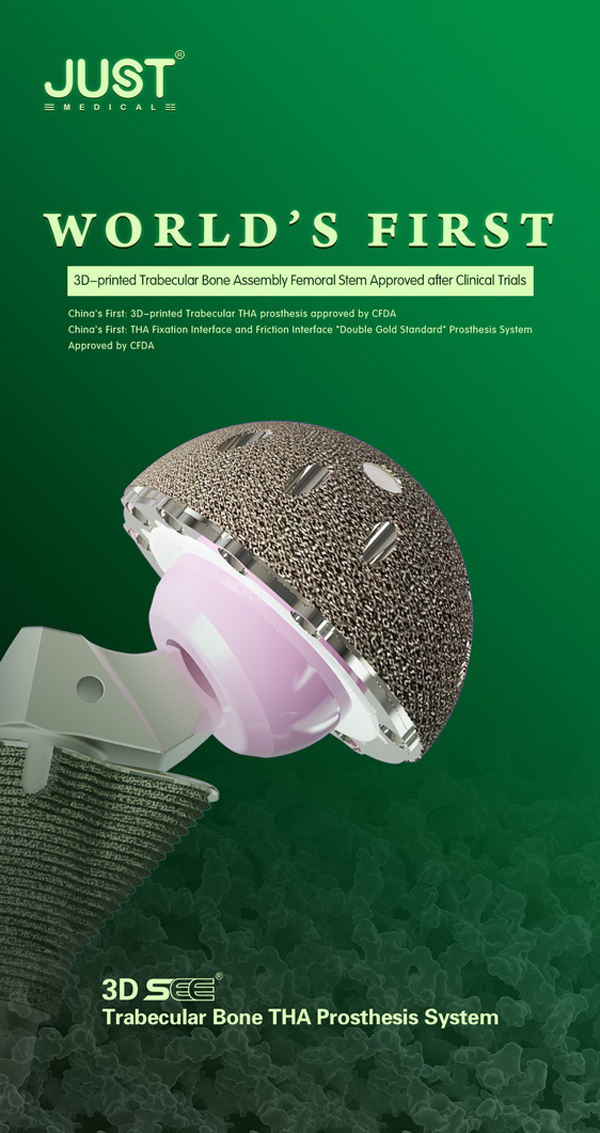
The product launch conference of 3D SEE® Trabecular Metal Hip Prosthesis System of JUST MED was successfully held in Xiamen, China on August 30, 2019! Four clinical investigators: Professor Fuxing Fu from West China Hospital of Sichuan University, Professor Wanshou Guo from China-Japan Friendship Hospital, Professor Zongke Zhou from West China Hospital of Sichuan University, and Professor Zhencai Shi from China-Japan Friendship Hospital introduced the clinical results at the main venue of the 2019 Chinese Joint Surgery Academic Conference in Xiamen. During the conference, relevant government leaders, clinicians and media representatives from all levels of hospitals attended the conference to witness the clinical research achievements of the world’s first 3D-printed trabecular modular stem.
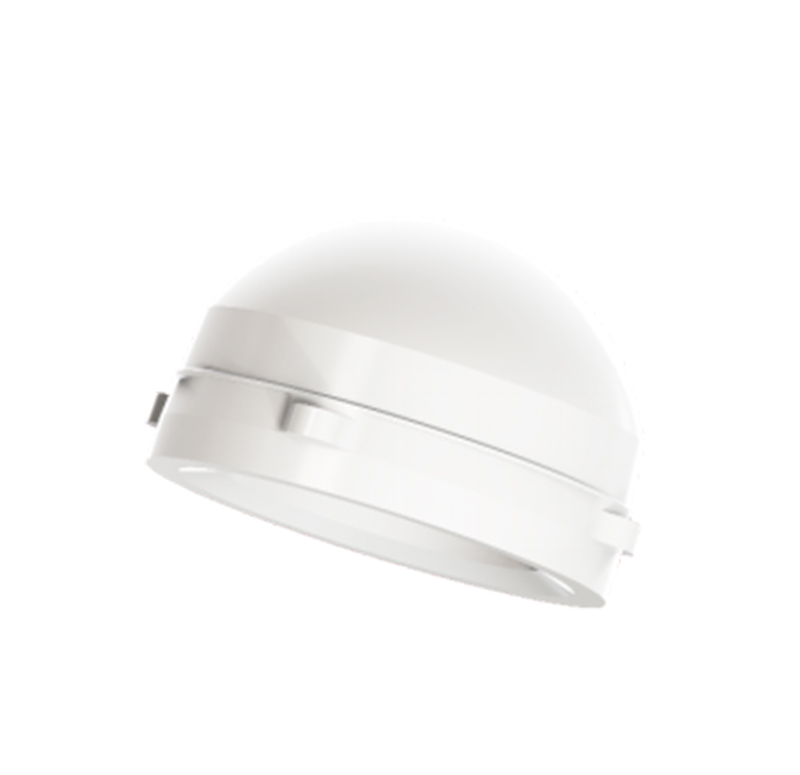
Hip Prosthesis Supplier
At the conference, four clinical researchers detailed the clinical research results of the 3D SEE® Trabecular Metal Hip Prosthesis System from four dimensions as why choose 3D printing trabecular bone modular femoral stem for clinical research, safety, effectiveness, and future prospects.
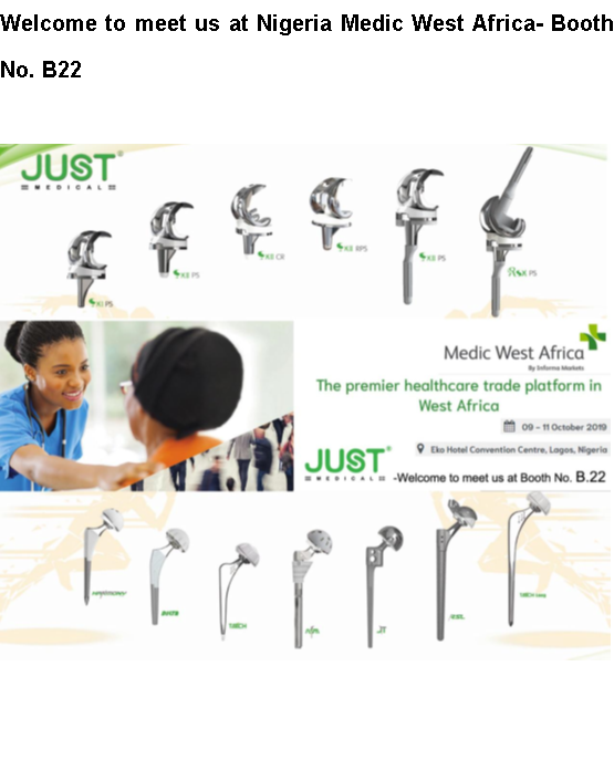
“Nigeria Medic West Africa” will be held in Africa from October 9 to October 11, 2019. As a professional Hip Prosthesis Accessories Manufacture, our company will take part in this exhibition. Warmly Welcome to join us and to learn more information about our product Hip Joint System Prosthesis.
The Nigeria Medic West Africa will be held in October 2019 with more than 4,500 attendees to meet the business needs of dealers and distributors, new customers and developments in West Africa and existing customers. Represented in 32 countries, participating in the largest commercial medical exhibition in West Africa, enhancing international influence and brand exposure, West Africa Nigeria Medical Exhibition is the largest commercial platform and a perfect exhibition for you to showcase your latest products and services to an active audience.
Hip Prosthesis Accessories Manufacture
The information about Nigeria Medic West Africa:
Time:2019.10.9-10.11
Address: Eko Convention Centre
Our company’s information:
Company Name: Just HuaJian Medical Device(TianJin)Co.,Ltd.
Booth No.: B22
We are a professional company who can supply Hip Joint Replacement. Hope to look for new business opportunities.
Amputees often wear prosthetic limbs. In order to ensure the normal use of the prosthetic and to play its functions and prolong the service life, Acetabular Cup Prosthesis Manufacturertells you the following items should be noted.
Hip Joint System Prosthesis Price
First, the formal acceptance of the cavity maintenance: (including the bushing silicone soft cover)
1. The upper limb arm and lower limb receiving cavity are kept clean every day, cleaned before going to bed, and dried naturally.
2. The upper limb arm and the lower limb receiving cavity sometimes wear residual limb atrophy phenomenon, at this time can be adjusted by wearing the residual body socks or filling the leather pad to keep wearing and walking safe. If you are still too loose, please contact the company in time to avoid wrestling after slipping, and replace the receiving cavity if necessary.
3. Silicone, gel soft cover wear notes: (use the correct guidance under the guidance of the technician)
(1) Cleaning method: Clean the inside with neutral soapy water daily and air dry. (Use a care solution is preferred).
(2) Avoid sharp and rough objects from being scratched and torn; avoid high temperature exposure and baking; avoid strong pull.
Second, temporary acceptance of the cavity and temporary prosthesis.
1. The temporary receiving cavity and the temporary prosthesis are used as an amputee for the initial prosthesis for observing, shaping and stabilizing the residual limb.
2. The temporary prosthesis is only used as the weight of the residual limb, support and joint mobility, training strength, and gait.
1. Warm-up activities before training must be sufficient. Do not directly exercise large amounts of exercise. The most common occurrence of injury is when fatigue or lack of energy.
2. Pay attention to the running posture when running, don’t just use one side of a foot to land;
3. Pay attention to the muscles of the thighs to avoid direct impact from the knees;
4. When there is a symptom of knee discomfort, properly reduce the amount of exercise and the frequency of exercise to avoid violent running, jumping and weight-bearing movements;
5. Pay attention to the knees to keep warm, especially in the summer, don’t be greedy;
6. Apply heat to the leg muscles after exercise;
7. When the weight is pulled, especially when going downhill, avoid the knee from being impacted;
8. The development of muscles can relieve the pressure on the knee under certain conditions, and can minimize the damage to the knee. Usually do more leg muscle exercise, try to reduce the resistance to the knee. Consciously strengthen the exercise of the quadriceps (anterior thigh) and cruciate ligament, strengthen the muscles of the quadriceps and thigh muscles (such as weight-bearing squats), combined with muscle stretching, walking, walking or sticking Wall practice and other methods, paving the way for the smooth movement of the tibia in the femur. I feel more relaxed when I go to Yuzhu this time.
9. Before climbing, use your hands and fingers to rub the lower edge of your knees to promote lubrication of your knees at night;
10. Stretching the elongated hamstrings and patellofemoral joints is beneficial to reduce the chance of knee injury.
It is the most important manifestation of arthritis. Different types of arthritis can exhibit different pain characteristics.
2. Joint swelling
Swelling is a common manifestation of joint inflammation and is the result of inflammation progression, not necessarily related to the degree of joint pain. Generally proportional to the disease.
3. Joint dysfunction
Joint edema around the joint caused by joint pain and inflammation, protective tendon of the surrounding muscles and joint structure are destroyed, resulting in limited joint activity. Chronic arthritis patients may have permanent joint function loss due to limited long-term joint activity.
Other symptoms
Different types of arthritis signs are also different, may occur erythema, deformity, soft tissue swelling, joint swelling, exudate, bone swelling, bone rubbing, tenderness, muscle atrophy or muscle weakness, limited range of joint activity and nerve root compression Equal signs.
Diagnose based on
1. Different types of arthritis have different signs, such as erythema, malformation, soft tissue swelling, joint swelling, exudate, bone swelling, bone rubbing, tenderness, muscle atrophy or muscle weakness, limited range of joint motion and nerve roots Signs such as pressure.
2. X-ray films of the joints can be seen soft tissue swelling, osteoporosis and subchondral bone sclerosis and/or articular surface cystic changes, invasive bone destruction, articular surface blur, asymmetrical joint space stenosis, joint fusion And dislocation.
3. Arthroscopy and joint synovial biopsy are valuable for the diagnosis and differential diagnosis of arthritis.
Treatment policy
Choose the appropriate treatment based on the type of arthritis, the characteristics of the symptoms, and the accompanying diseases. The principle of treatment is early diagnosis and rational and combined use as soon as possible.
medical treatement
Commonly used anti-rheumatic drugs are: non-steroidal anti-inflammatory drugs ibuprofen, penicillamine, diclofenac, aspirin, indomethacin and so on. Chondroprotective agent glucosamine sulfate. Slow-acting anti-rheumatic drug gold mixture (intramuscular or oral), penicillamine, sulfasalazine, chloroquine, and the like. Cytotoxic drugs cyclophosphamide, methotrexate, Jin Duchun and so on. Adrenocorticotropic hormone. Antibiotics, anti-tuberculosis or antifungal drugs.
Treatment for gouty arthritis includes high-dose non-steroidal anti-inflammatory drugs or colchicine and uric acid-lowering therapy during remission.
Surgical treatment
Surgical treatment mainly includes joint cavity puncture, synovectomy, joint replacement, joint orthopedics, joint fusion and so on.
There are many reasons for hip pain, which is simply classified as:
First, traumatic factors such as trauma, joint pain caused by falls.
Second, the factors of degeneration, such as age, the deformation of the tissues in and around the joints, may cause joint pain.
Third, the systemic immune factors, in front of the hip joint.
Fourth, it is the corresponding blood supply factors of the joint, such as common clinical disease, avascular necrosis of the femoral head, etc,.
Fifth, there are some other factors, such as The lumbar spine is expressed on the hip joint or is a systemic tuberculosis or a local tumor lesion, which is manifested in the hip joint.
As a Joint Prosthesis Supplier, today we like to share with you the following knowledge, which can help you to learn the treatment of residual limb care and complications after amputation.
Good residual limbs are a prerequisite for the assembly of prostheses, and the amputation and amputation should be considered during surgery. The following are treatments for post-operative residual limb care and its complications:
First, maintain a good position.
Prevent joint contracture and deformity of residual limbs.
As part of the muscle is cut off after amputation, it will cause muscle imbalance and joint contracture. Such as: hip flexion, hip abduction, knee flexion, ankle joint plantar flexion, the results will affect the alignment of the prosthesis. Therefore, postoperative should be contraindicated: raise the affected area when lying on the back, pad the pillow in the perineum and make the thigh abduction, long-term wheelchair ride, lift the limb with a wooden turn and other bad posture. Postoperative joints should be placed in the functional position, and early functional exercise to make the joints flexible and deformed.
Second, slow down the pain of residual limbs: there are many reasons for pain, mainly in the following aspects:
(a) surgery for nerve stimulation
(b) residual blood circulation disorder
(c) abnormal muscles in which the residual limb is severed
(d) residual limbs have neuroma
We are selling kinds of joint prosthesis, if you have any question about Joint Prosthesis Price, feel free to let us know.
Here is a Knee Joint Preservation Manufacturer talking about knee training. If you have any idea about it, you can contact us and share it with us. Also, our company has various Knee Prosthesis Types. If you need or you are interested in it, feel free to let us know at any time. Now let us learn some training methods of knee joint below.
Knee Prosthesis Types
The knee joint belongs to the hinge joint and it can only move forward and backward. In the case that the knee joint has been injured, first of all, pay attention to protection. If possible, you can perform simple back and forth movement to promote blood circulation of the knee joint, but it is not suitable for strong activity. The exercise is not easy enough to cause stiffness of the knee joint. Excessive movement will accelerate the wear and tear of the joint, but the movement still does not feel pain in the joints.
Preventive health care of the knee joint through proper muscle strength and stability training:
1. Lying on the side: lying on the left side, slightly bent on the knees, and the heels are close together. The headrest is on the left arm and the eyes are looking straight ahead. Hold a weight of about 1 to 2 kilograms on the right and place it on the outside of the leg. Then the abdomen is tightened, the buttocks are tight, and the knees of the right leg are raised as much as possible. When the legs are lifted, the body does not move, hold for a few seconds, and let go. Repeat the exercise 15 times and change legs.
2. Leg raising exercises: stand behind a stable bench or step, the right foot stepped on (the heel should not be suspended), and the weight is concentrated on the right foot, the body is raised, the left foot toe touch the step, adhere to 1 to 5 seconds bell. Then lower your left foot and slap the ground. Repeat 8 to 10 times and change legs.
3. Bridging practice: lying flat on the ground, knees bent, feet apart, with the hip width, arms on both sides. Slowly lift your hips and leave the ground smoothly. Then slowly put it down. Repeat 15 times.
4. Leg movement: lying flat, knees bent, feet flat on the ground. Extend the left leg and fit into the telescopic
Pull the belt or towel and grasp the ends of the strap with both hands. Pull the leg to the chest with a strap and force the calf straight for 10 to 30 seconds to exercise the calf muscles and tendons. Repeat the action 3 to 5 times and then change legs.
Here we are talking about hip pain, also we are a factory who sells Hip Replacement. If you have any question about Hip Prosthesis Price, you can contact us.
Hip Replacement
There are three common reasons why there exists hip pain:
1. It is generally believed that the cause of hip joint pain in people with normal hip joint structure is roughly divided into hip arthritis lesions and lesions of the femoral head itself, which may often exist at the same time. Hip arthritic lesions can be further divided into lesions caused by bacterial infections and aseptic inflammation caused by no bacteria. Hip arthritis with bacterial infection can be caused by Mycobacterium tuberculosis or septic arthritis caused by other bacteria. Articular cartilage must be destroyed and the prognosis is often poor. The most common form of aseptic inflammation is “synovitis”. This type of inflammation may be caused by trauma or impact on the joints. It is usually transient, and you can recover by yourself after a period of rest. The problem is not big. Another type of aseptic inflammation is rheumatoid factor, rheumatoid factor or synovitis secondary to femoral head necrosis. This type of synovitis may have a serious prognosis.
2. There is also a type of patient with hip joint pain due to abnormal hip structure. Such patients may have a special history of hip joints, such as abnormal development of the hip joint structure or a history of hip dislocation, etc., often with occasional joint pain, the general situation can be improved by rest and taking some anti-inflammatory analgesics, the problem is not big . If the joint is painful and cannot be improved after taking the medicine and rest, consult a doctor to evaluate further treatments, reduce and treat hip pain.
3. The third type is femoral head necrosis, which is also the most common and most harmful type of disease in the hip joint disease spectrum. About 200,000 people are sick each year. Because newspapers and advertisements often refer to this disease, when the patient has pain in the hip joint, he or she often causes panic because of fear of the disease. However, the primary symptom of femoral head necrosis is hip pain.
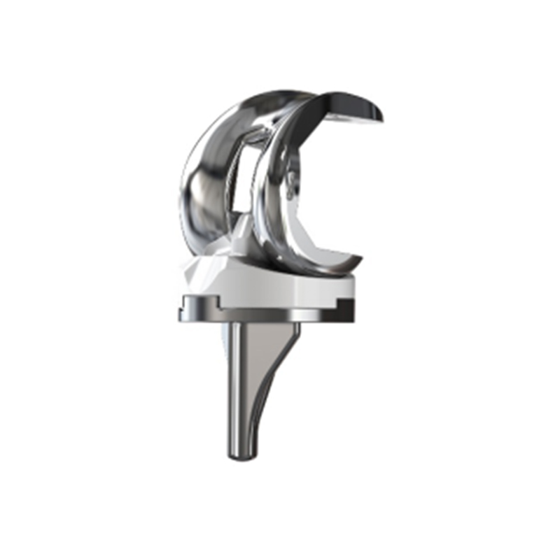
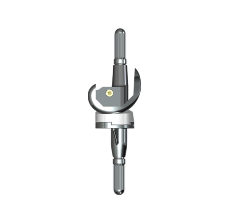
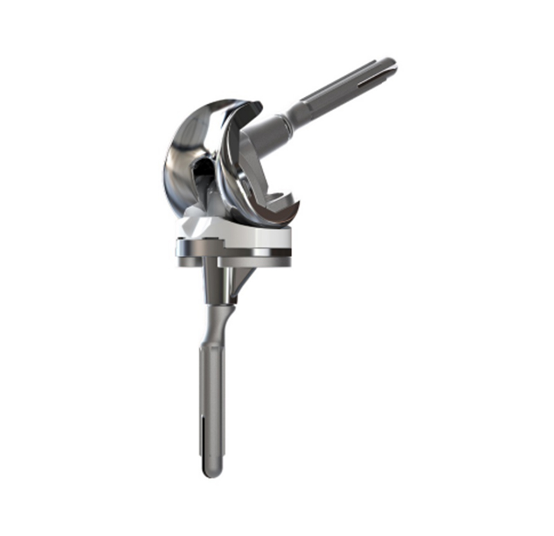
The knee joint is the largest and most complex joint in the human body, including three bones: the femur (thigh bone), the tibia (calf bone) and the tibia (knee bone). In daily life, walking, walk up and down stairs, sitting up play an important role in our life. If the articular surface of the knee joint wears is damaged or destroyed due to various reasons, resulting in narrow joint space, walking pain and dysfunction, it will seriously affect the quality of life. Artificial knee replacement is one of the most effective and reliable methods for the treatment of advanced knee arthritis. Revision Knee Joint Prosthesis is also a good way to cure it.
However, for an elderly patient, artificial knee arthroplasty is not a non-invasive or minimally invasive procedure like a denture. It requires a high level of preoperative preparation. The correct prosthesis selection is to ensure satisfactory surgical outcome.
Revision Knee Joint Prosthesis
At present, there are many types of knee prostheses commonly used in clinical practice, and there are more than a dozen manufacturers. As a professional Revision Knee Prosthesis Manufacturer, today we understand the composition of the prosthesis.
The knee prosthesis consists of the following three parts:
1. Femoral prosthesis: the surface replaces the end of the femur. The femoral prosthesis consists of a metal alloy.
2. Humeral prosthesis: instead of the inner surface of the tibia rubbing with the femur. The tibial prosthesis consists of a plastic with a metal alloy backing.
3. Humeral prosthesis: it can be single or two designs. The single design consists of plastic and the two designs consist of a metal holder and a plastic sheet attached to the bone. The plastic spacer provides a smooth surface on which the femur moves. The plastic spacer is usually attached to the tibial tray.